Your How does fluorescence microscopy work images are ready in this website. How does fluorescence microscopy work are a topic that is being searched for and liked by netizens today. You can Find and Download the How does fluorescence microscopy work files here. Download all royalty-free photos and vectors.
If you’re searching for how does fluorescence microscopy work images information connected with to the how does fluorescence microscopy work interest, you have visit the right site. Our site frequently gives you suggestions for seeking the maximum quality video and picture content, please kindly hunt and locate more enlightening video articles and images that fit your interests.
How Does Fluorescence Microscopy Work. How does fluorescence imaging work. The specimen is illuminated by light with a specific wavelength that is absorbed by the fluorophores enabling them to emit light of longer. A fluorescence microscope works by combining the magnifying properties of the light microscope with fluorescence emitting properties of compounds. A majority of the excitation light reaching the specimen passes through without interaction and travels away from the objective and the illuminated area is restricted to.
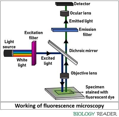 What Is Fluorescence Microscopy Definition Principle Fluorescence Parts Biology Reader From biologyreader.com
Fluorescence is the emission of light by a substance that has absorbed light or other electromagnetic radiation while phosphorescence is a. When these probes are applied a fluorescent microscope can be used to detect the presence or absence of individual microbial groups. How Does Fluorescence Microscopy Work. The basic fluorescence microscope is a common tool of modern cell biologists. Ad Live-Cell the Fixed-Cell Compatible. How does Palm microscopy work.
Fluorescence microscopy is a particular form of light microscopy in which instead of utilizing visible light to illuminate specimens a higher intensity light source excites a fluorescent molecule called a fluorophore.
The specimen is illuminated by light with a specific wavelength that is absorbed by the fluorophores enabling them to emit light of longer. Images are formed by the simultaneous observation of many fluorescing molecules to visualize cellular structures and processes. Fluorescent probes are designed to attach to specific genetic regions of microbes that will differentiate them from other groups. Fluorescence microscopy is a type of light microscope that works on the principle of fluorescence. A fluorescence microscope uses a mercury or xenon lamp to produce ultraviolet light. How does confocal microscopy work.
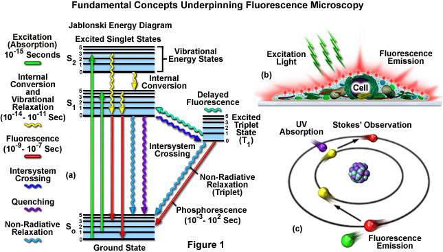 Source: zeiss-campus.magnet.fsu.edu
How Does Fluorescence Microscopy Work. A fluorescence microscope is an optical microscope that uses fluorescence and phosphorescence instead of or in addition to reflection and absorption to study properties of organic or inorganic substances. In conventional far-field fluorescence microscopy the. Once we know what is fluorescence the principle of fluorescence microscope becomes simple. How does Palm microscopy work.
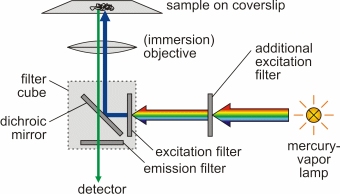 Source: home.uni-leipzig.de
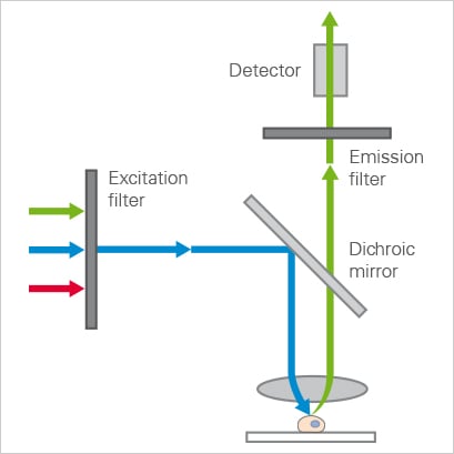
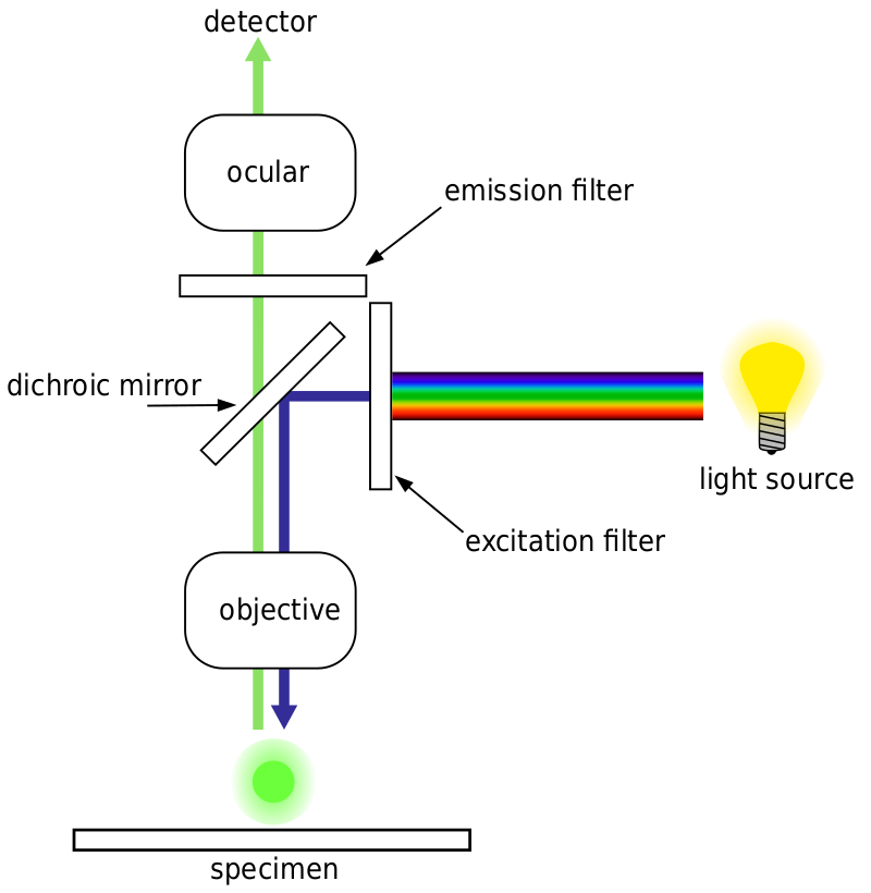
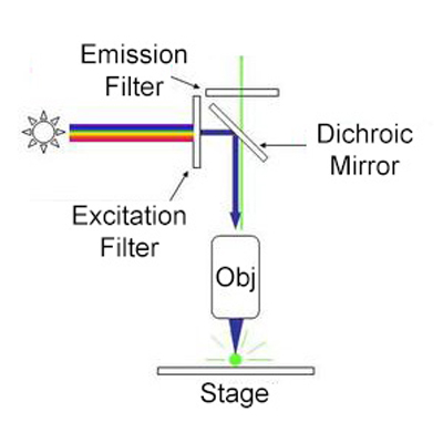
The light comes into the microscope and hits a dichroic mirror – a mirror that reflects one range of wavelengths and allows another range to pass through. The ultraviolet light emitted from the microscope is reflected by. The basic fluorescence microscope is a common tool of modern cell biologists. How does fluorescence imaging work. Fluorescence microscopy is a type of light microscope that works on the principle of fluorescence.

The ultraviolet light emitted from the microscope is reflected by. Fluorescence microscopy is a type of light microscope that works on the principle of fluorescence. This mirror reflects light shorter than a certain wavelength and passes light longer than that. Fluorescence is the emission of light that occurs within nanoseconds after the absorption of light that is typically of. How does fluorescence microscopy work.
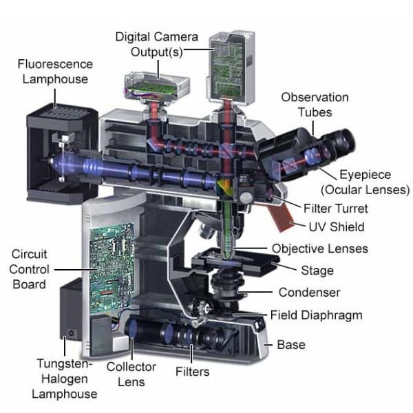 Source: microscopeinternational.com
The microscope uses a special dichroic mirror or more properly a dichromatic mirror although this term only seems to be used by purists. When these probes are applied a fluorescent microscope can be used to detect the presence or absence of individual microbial groups. Immediate Imaging or for Long-Term Storage. Fluorescent probes are designed to attach to specific genetic regions of microbes that will differentiate them from other groups. How does confocal microscopy work.
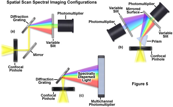 Source: zeiss-campus.magnet.fsu.edu
Ad Live-Cell the Fixed-Cell Compatible. Ad Live-Cell the Fixed-Cell Compatible. The microscope uses a special dichroic mirror or more properly a dichromatic mirror although this term only seems to be used by purists. After fluorophores are applied to the sample in the area of interest the sample is exposed to electromagnetic energy at a certain wavelength which excites the fluorophores to. Fluorescence microscopy requires that the objects of interest fluoresce.
 Source: ibidi.com
The microscope uses a special dichroic mirror or more properly a dichromatic mirror although this term only seems to be used by purists. Fluorescence is the emission of light that occurs within nanoseconds after the absorption of light that is typically of. Traditionally excitation light is provided by a mercury or xenon high-pressure bulb and the. Refractive Index of 152. Do surgeons use virtual reality.
 Source: sciencedirect.com
Do surgeons use virtual reality. The ultraviolet light emitted from the microscope is reflected by. In order to detect the fluorescent light using an optical microscope there are three steps. How Does Fluorescence Microscopy Work. Immediate Imaging or for Long-Term Storage.
 Source: semrock.com
In this instrument a parallel beam of light simultaneously illuminates the whole specimen or wide-field of view to excite via the filter block the fluorophores it contains. Fluorescence microscopy uses a high-intensity light source that excites a fluorescent molecule called a fluorophore in the sample observed. Immediate Imaging or for Long-Term Storage. Fluorescence is the emission of light by a substance that has absorbed light or other electromagnetic radiation while phosphorescence is a. A fluorescence microscope works by combining the magnifying properties of the light microscope with fluorescence emitting properties of compounds.
 Source: pinterest.com
How does fluorescence microscopy work. Once we know what is fluorescence the principle of fluorescence microscope becomes simple. A fluorescence microscope uses a mercury or xenon lamp to produce ultraviolet light. It is an optical technique that relies on fluorescent light as the light source. The microscope uses a special dichroic mirror or more properly a dichromatic mirror although this term only seems to be used by purists.
 Source: youtube.com
Fluorescence microscopy uses a high-intensity light source that excites a fluorescent molecule called a fluorophore in the sample observed. The dichroic mirror reflects the ultraviolet light up to the specimen. In conventional far-field fluorescence microscopy the. In this animation you will be introduced to fluorescence microscopy which is a specialized type of light microscopy. The ultraviolet light emitted from the microscope is reflected by.
 Source: photometrics.com
Fluorescence is the emission of light that occurs within nanoseconds after the absorption of light that is typically of. The light comes into the microscope and hits a dichroic mirror – a mirror that reflects one range of wavelengths and allows another range to pass through. A fluorescence microscope works by combining the magnifying properties of the light microscope with fluorescence emitting properties of compounds. When these probes are applied a fluorescent microscope can be used to detect the presence or absence of individual microbial groups. How does Palm microscopy work.
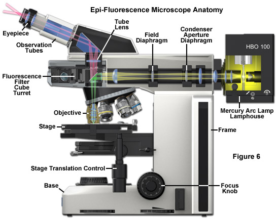 Source: zeiss-campus.magnet.fsu.edu
Fluorescence microscopy is a popular imaging technique both for its relatively noninvasive properties and its ability to simultaneously image multiple species in living cellular specimens. Fluorescence microscopy is a popular imaging technique both for its relatively noninvasive properties and its ability to simultaneously image multiple species in living cellular specimens. Ad Live-Cell the Fixed-Cell Compatible. Fluorescence microscopy is a particular form of light microscopy in which instead of utilizing visible light to illuminate specimens a higher intensity light source excites a fluorescent molecule called a fluorophore. How Does Fluorescence Microscopy Work.
 Source: biologyreader.com
Fluorescence microscopy is a popular imaging technique both for its relatively noninvasive properties and its ability to simultaneously image multiple species in living cellular specimens. This mirror reflects light shorter than a certain wavelength and passes light longer than that. When you hear the word microscope chances are what first comes to mind is actually fluorescence microscopy. The dichroic mirror reflects the ultraviolet light up to the specimen. How does Palm microscopy work.
 Source: microscopeinternational.com
In this instrument a parallel beam of light simultaneously illuminates the whole specimen or wide-field of view to excite via the filter block the fluorophores it contains. Images are formed by the simultaneous observation of many fluorescing molecules to visualize cellular structures and processes. How does a fluorescence microscope work. The basic fluorescence microscope is a common tool of modern cell biologists. When you hear the word microscope chances are what first comes to mind is actually fluorescence microscopy.
 Source: researchgate.net
In this animation you will be introduced to fluorescence microscopy which is a specialized type of light microscopy. Traditionally excitation light is provided by a mercury or xenon high-pressure bulb and the. A fluorescence microscope works by combining the magnifying properties of the light microscope with fluorescence emitting properties of compounds. Immediate Imaging or for Long-Term Storage. In this instrument a parallel beam of light simultaneously illuminates the whole specimen or wide-field of view to excite via the filter block the fluorophores it contains.
 Source: sciencedirect.com
How does a fluorescence microscope work. The specimen is illuminated by light with a specific wavelength that is absorbed by the fluorophores enabling them to emit light of longer. Immediate Imaging or for Long-Term Storage. A majority of the excitation light reaching the specimen passes through without interaction and travels away from the objective and the illuminated area is restricted to. When these probes are applied a fluorescent microscope can be used to detect the presence or absence of individual microbial groups.
 Source: home.uni-leipzig.de
Fluorescence microscopy is a popular imaging technique both for its relatively noninvasive properties and its ability to simultaneously image multiple species in living cellular specimens. How does fluorescence imaging work. In conventional far-field fluorescence microscopy the. Fluorescence microscopy requires that the objects of interest fluoresce. Being a single component the objectivecondenser is always in perfect alignment.
 Source: rsscience.com
Do surgeons use virtual reality. How does fluorescence microscopy work. After fluorophores are applied to the sample in the area of interest the sample is exposed to electromagnetic energy at a certain wavelength which excites the fluorophores to. A majority of the excitation light reaching the specimen passes through without interaction and travels away from the objective and the illuminated area is restricted to. In the picture above we are supposing the excitation light needs to be violet and the emitted light is red.
This site is an open community for users to submit their favorite wallpapers on the internet, all images or pictures in this website are for personal wallpaper use only, it is stricly prohibited to use this wallpaper for commercial purposes, if you are the author and find this image is shared without your permission, please kindly raise a DMCA report to Us.
If you find this site adventageous, please support us by sharing this posts to your own social media accounts like Facebook, Instagram and so on or you can also bookmark this blog page with the title how does fluorescence microscopy work by using Ctrl + D for devices a laptop with a Windows operating system or Command + D for laptops with an Apple operating system. If you use a smartphone, you can also use the drawer menu of the browser you are using. Whether it’s a Windows, Mac, iOS or Android operating system, you will still be able to bookmark this website.

